By: Jeb Helms, PT, DPT, OCS, SCS
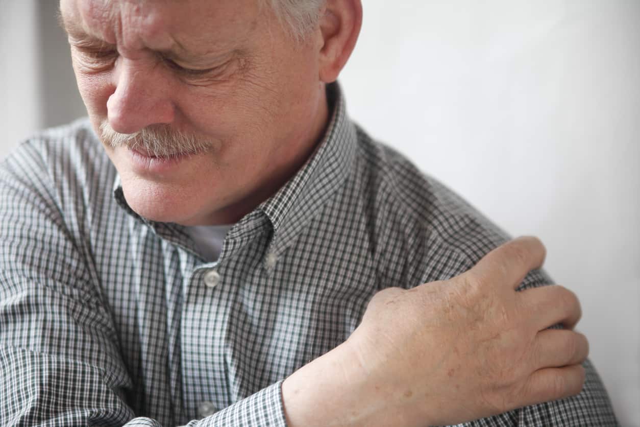 Imagine this all too common PT, OT, or AT patient scenario: You’re evaluating a patient with shoulder pain, and every shoulder motion, special test, or strength assessment reproduces pain not just at their shoulder but in other areas of the body. Additionally, your patient is convinced the arthritis in their shoulder that they saw on a recent X-ray only partly explains their continued pain. They ask you why they are seeing you as a rehab professional and not getting an MRI to find out “What’s really wrong”. Or maybe they had that MRI and just found out that in addition to arthritis, they have tears in their labrum and rotator cuff and are frustrated they are sitting with you and not undergoing surgery right away.
One of the challenges for us as rehab professionals is shifting our mindset as clinicians from a belief that all pain has a distinct anatomical cause towards an understanding that many things play into a person’s pain experience. I know for me as a student, I learned my “rotator cuff special tests” and “labrum special tests” but would often be confused when their results did not match up with patients’ imaging results. As I became more aware of the many studies showing normal pain-free patients with spinal disc problems at the cervical spine (Nakashima et al., 2015) and lumbar spine (Baker, 2014) as well as hip labral tears (Tresch et al., 2017), knee OA (Culvenor et al., 2019), and rotator cuff tears (Tempelhof et al., 1999), I struggled over how to convey that information to my patients. I’d want to tell my patients “Yes, you’re in pain and yes your MRI showed something wrong with your shoulder but did you know that your other shoulder that doesn’t hurt likely has a similar looking MRI as the one you’re here to see me for (Barreto et al., 2019)? Or that successful rotator cuff surgery repairs often re-tear by a 10-year follow-up but patients are still pain-free and happy with their repair (Spielmann et al., 1999). I might even touch on intriguing research around placebo surgeries at the knee (Sihvonen et al., 2018) and shoulder (Paavola et al., 2018) that point to the power of patient expectations in influencing pain.
Chester et al. (2018) showed that shoulder ROM, strength, and special test assessment could not accurately predict how a patient would do functionally 6 months later. Instead, psychological variables like patient expectations of improvement were much more predictive. Flynn et al. (2011) accurately point out that imaging results are often more harmful than helpful in shaping patient expectations around recovery. To combat that fact, we as rehab professionals need to be able to easily explain imaging results to patients in 3 distinct steps:
- Listen to our patients tell their story to build a therapeutic alliance. Diener et al. (2016) lay out a great framework on how to do this clinically. Without patients feeling like you listened to their story, any imaging education will likely fall on deaf ears.
- Discuss research around pain-free imaging findings. For the low back, this systematic review is a great place to start (Brinjikji et al., 2015). Our clinic even made posters with the results of this study to place in the waiting room to start conversations with our patients.
- Raise patient expectations around the imaging results. Point out what the imaging doesn’t show, like a fracture for example. “Yes, you have a partial rotator cuff tear but that’s normal for someone your age, I’m just really glad that it didn’t show a fracture or a complete tear of your cuff – I really think I should be able to help you. I also love using symptom modification tests as well to demonstrate how pain might change during the exam downplaying the results of the imaging.
What do you think? How do you approach imaging findings when working with a patient?
Interested in learning more about pain assessment and treatment? We have several courses that may help. Course topics include Pain Science courses on evaluation, general treatment, manual therapy, and, coming soon, difficult pain conditions like CRPS that will help give you a plan on clinical uses of pain science in the clinic. Check them out below. Also, if you are interested in all 4 of our current offerings we will take 20% off if you purchase all 4 together! Just enter the coupon code “PAIN20” at checkout and save.
Chronic Pain & Pain Neuroscience Education for the PT, OT, & AT: The Evaluation
Chronic Pain & Pain Neuroscience Education for the PT, OT, & AT: Treatment
Manual Therapy from a Pain Neuroscience Education Approach
References
Baker, A. D. (2014). Abnormal magnetic-resonance scans of the lumbar spine in asymptomatic subjects. A prospective investigation. In Classic papers in orthopaedics (pp. 245-247). Springer. https://doi.org/10.2106/00004623-199072030-00013
Barreto, R. P. G., Braman, J. P., Ludewig, P. M., Ribeiro, L. P., & Camargo, P. R. (2019). Bilateral magnetic resonance imaging findings in individuals with unilateral shoulder pain. Journal of shoulder and elbow surgery, 28(9), 1699-1706. https://doi.org/10.1016/j.jse.2019.04.001
Brinjikji, W., Luetmer, P. H., Comstock, B., Bresnahan, B. W., Chen, L. E., Deyo, R. A., Halabi, S., Turner, J. A., Avins, A. L., James, K., Wald, J. T., Kallmes, D. F., & Jarvik, J. G. (2015). Systematic literature review of imaging features of spinal degeneration in asymptomatic populations. AJNR. American journal of neuroradiology, 36(4), 811-816. https://doi.org/10.3174/ajnr.A4173
Chester, R., Jerosch-Herold, C., Lewis, J., & Shepstone, L. (2018). Psychological factors are associated with the outcome of physiotherapy for people with shoulder pain: A multicentre longitudinal cohort study. British journal of sports medicine, 52(4), 269-275. https://doi.org/10.1136/bjsports-2016-096084
Culvenor, A. G., Øiestad, B. E., Hart, H. F., Stefanik, J. J., Guermazi, A., & Crossley, K. M. (2019). Prevalence of knee osteoarthritis features on magnetic resonance imaging in asymptomatic uninjured adults: a systematic review and meta-analysis. British journal of sports medicine, 53(20), 1268-1278. https://doi.org/10.1136/bjsports-2018-099257
Diener, I., Kargela, M., & Louw, A. (2016). Listening is therapy: Patient interviewing from a pain science perspective. Physiotherapy Theory & Practice, 32(5), 356-367. https://doi.org/10.1080/09593985.2016.1194648
Flynn, T. W., Smith, B., & Chou, R. (2011). Appropriate use of diagnostic imaging in low back pain: A reminder that unnecessary imaging may do as much harm as good. Journal of Orthopaedic & Sports Physical Therapy, 41(11), 838-846. https://doi.org/10.2519/jospt.2011.3618
Nakashima, H., Yukawa, Y., Suda, K., Yamagata, M., Ueta, T., & Kato, F. (2015). Abnormal findings on magnetic resonance images of the cervical spines in 1211 asymptomatic subjects. Spine, 40(6), 392-398. https://doi.org/10.1097/BRS.0000000000000775
Paavola, M., Malmivaara, A., Taimela, S., Kanto, K., Inkinen, J., Kalske, J., Sinisaari, I., Savolainen, V., Ranstam, J., & Järvinen, T. L. (2018). Subacromial decompression versus diagnostic arthroscopy for shoulder impingement: Randomised, placebo surgery controlled clinical trial. Bmj, 362, k2860. https://doi.org/10.1136/bmj.k2860
Sihvonen, R., Paavola, M., Malmivaara, A., Itälä, A., Joukainen, A., Nurmi, H., Kalske, J., Ikonen, A., Järvelä, T., & Järvinen, T. A. (2018). Arthroscopic partial meniscectomy versus placebo surgery for a degenerative meniscus tear: A 2-year follow-up of the randomised controlled trial. Annals of the rheumatic diseases, 77(2), 188-195. https://doi.org/10.1136/annrheumdis-2017-211172
Spielmann, A. L., Forster, B. B., Kokan, P., Hawkins, R. H., & Janzen, D. L. (1999). Shoulder after rotator cuff repair: MR imaging findings in asymptomatic individuals—initial experience. Radiology, 213(3), 705-708. https://doi.org/10.1148/radiology.213.3.r99dc09705
Tempelhof, S., Rupp, S., & Seil, R. (1999). Age-related prevalence of rotator cuff tears in asymptomatic shoulders. Journal of shoulder and elbow surgery, 8(4), 296-299. https://doi.org/10.1016/S1058-2746(99)90148-9
Tresch, F., Dietrich, T. J., Pfirrmann, C. W., & Sutter, R. (2017). Hip MRI: Prevalence of articular cartilage defects and labral tears in asymptomatic volunteers. A comparison with a matched population of patients with femoroacetabular impingement. Journal of Magnetic Resonance Imaging, 46(2), 440-451. https://doi.org/10.1002/jmri.25565